BIOLOGY 70
INTRODUCTION TO PSYCHOBIOLOGY
LECTURES 2-3
back to index
TOPICS: READING: FIGURES FOR LECTURES 2-3:
Relevant WWW links:
PowerPoint Handouts FOR LECTURE 2-3: Lecture 2-3 PowerPoint Handouts(PDF)
Handouts without color backgrounds (PDF)
Luminance gradient
Craik-Obrien (Bach)
Craik-Obrien (Duke)

OVERVIEW: Once an image has been formed on the retina and visual transduction has occurred, neurons in the retina and the brain are ready to begin some serious information processing. In these lectures we will first discuss some perceptual phenomena related to the functioning of receptors. A second, major, aim of this section will be to see how interactions among neurons lead to transformation of the original “photograph” into new codes which emphasize certain aspects of the image while discarding others. We will discuss how this code is refined as information is transmitted along pathways to the brain.
- What are the differences between the rod and cone receptors with respect to:
a. numerosity d. color vision b. distribution across the retina e. visual resolution c. scotoptic and photopic vision - Know the following terms associated with the cells of the retina and retinal structure:
a. rods e. amacrine cells b. cones f. ganglion cells c. horizontal cells g. ribbon synapse d. bipolar cells h. optic nerve - What are the synaptic connections among the cells of the retina? What kinds of information are coded by each cell type (very generally)? In vertebrates, do receptors hyperpolarize or depolarize in response to light? (See figures 6.2 and 6.15 in Kalat and figure in “figures for lectures 2-3”.)
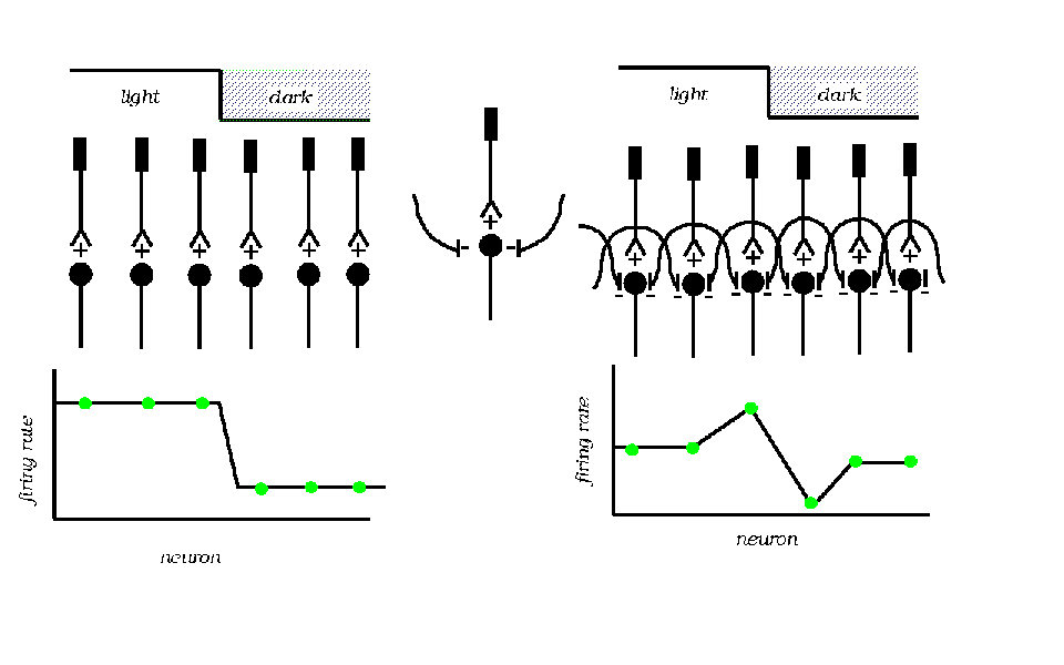
- Lateral inhibition is an important example of coding by neural networks.
Be sure to understand the discussion on pp. 167-169 in Kalat and the limulus evidence pictured in the “Lateral Inhibition” figure from Scientific American reproduced in “figures for lectures 2-3”. Also the diagram used in class.
- Understand how the following psychophysical phenomena are related to processes occurring in the retina:
- dark adaptation
- Pulfrich pendulum
- Mach bands
- Know the following terms related to the gross anatomy of the central visual system and their general function in visual information processing.
a. optic nerve f. inferior temporal cortex b. optic chiasm g. medial temporal cortex (MT, V5) c. lateral geniculate nucleus (LGN) and medial superior cortex (MST) d. superior colliculus h. ventral (temporal cortex) vs. dorsal (parietal cortex) streams e. visual cortex (V1, V2, V4) i. fusiform area - Understand the following functional concepts:
a. receptive field g. simple cell b. retinotopic map h. complex cell c. feature detector i. "grandmother" cell d. concentric on-center receptive field j. spatial frequency detector e. concentric off-center receptive field k. what vs where pathways f. orientationally tuned neuron - What does the Craik-Obrien illusion imply about information processing by the visual system?
- In the "simple" picture what are the types of information selectively processed by the parvocellular and magnocellular pathways (pp. 171-172, Table 6.2 and Figure 6.19 of Kalat)?
- Compare the "classical feature" and "spatial frequency" models of visual image processing.
- How is psychophysical adaptation used to show feature selectivity in the Blakemore-Sutton demonstration (see Figure in “figures for lectures 2-3” and WWW demo) and the McCulloch effect?
- What types of information are processed by the ventral (temporal) and dorsal (parietal) cortical streams?
- What is blindsight and which visual pathway may be implicated?